Navigating the world of ultrasounds can be overwhelming, especially for expectant parents eager to understand their baby’s development.
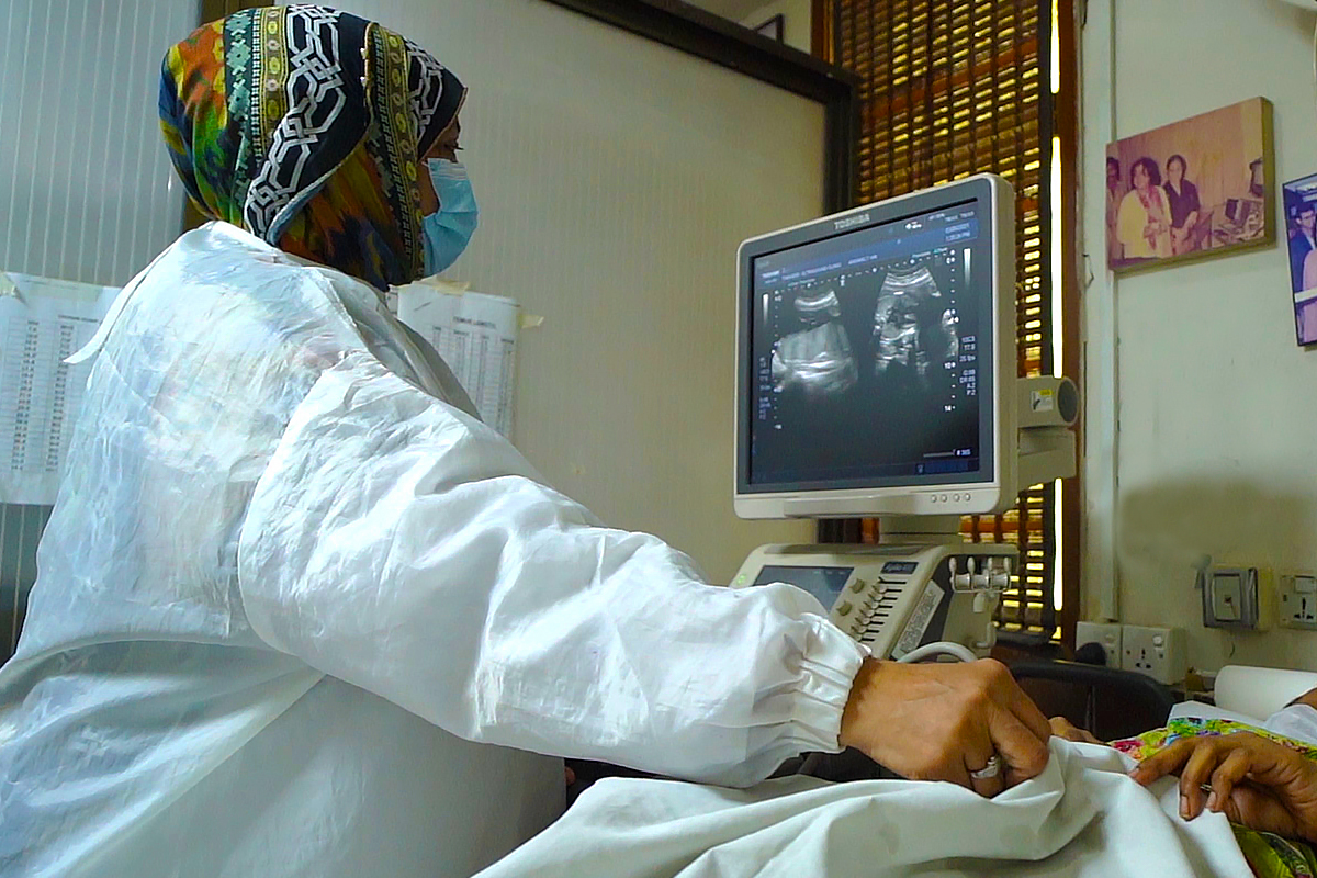
At Tanveer Ultrasound Clinic, we had the opportunity to sit down with Dr. Raheela, who shed light on the essential differences between 2D and 3D ultrasounds. With a gentle approach, she addressed common concerns about safety and the specific insights each type of ultrasound offers.
During our conversation, Dr. Raheela illuminated the key differences between 2D and 3D Ultrasounds with reassuring clarity.
“2D ultrasounds,” she explained, “are the traditional black-and-white images most people are familiar with. These images are essentially slices of the body, providing a flat, two-dimensional view of the fetus.”
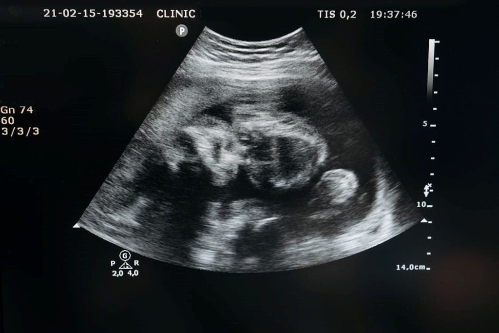
A 2D ultrasound offers a flat, functional view of the baby.
This method is particularly effective for routine checks, measuring fetal growth, congenital anomalies and monitoring the baby’s heartbeat.
3D Ultrasound: A Detailed, Lifelike Perspective
In contrast, 3D ultrasounds offer a more detailed and lifelike image.
“With 3D ultrasounds,” Dr. Raheela noted, “we can capture volumetric data, which allows us to construct a three-dimensional image. This is incredibly useful for visualizing the baby’s facial features and can provide a more comprehensive view of structural anomalies.”
Throughout our discussion, Dr. Raheela emphasized that both types of ultrasounds have their unique strengths and are chosen based on the specific needs of the patient.
At Tanveer Ultrasound Clinic, the choice between 2D and 3D is carefully considered to provide the best care and most accurate information possible.
“Timing is essential when it comes to choosing between 2D and 3D ultrasounds.” – Dr. Raheela
“We typically perform 2D ultrasounds throughout the entire pregnancy, as they are invaluable for regular monitoring of fetal growth, checking the baby’s heartbeat, and conducting anomaly scans. These scans are usually done between 20 to 22 weeks and provide critical insights into the baby’s development and any potential issues.”
As we delved deeper into the topic, Dr. Raheela elaborated on the timing for 3D ultrasounds.
“While 3D ultrasounds can technically be performed at any stage, we often recommend them between 24 to 32 weeks. This is because, during this period, there’s enough amniotic fluid and the baby has developed sufficiently, allowing us to capture more detailed and clearer images. It’s especially useful for parents eager to see their baby’s facial features or for medical professionals needing a more detailed view of any detected anomalies.”
Safety and Expertise at Tanveer Ultrasound Clinic
Addressing the safety concerns that many expectant parents have, Dr. Raheela reassured us, “It’s completely understandable for parents to worry about the safety of ultrasounds, but I want to emphasize that both 2D and 3D ultrasounds are generally very safe.”
She explained that ultrasounds use sound waves to create images, and unlike X-rays, they do not involve ionizing radiation. This makes them a safer option for both mother and baby.
“We follow strict guidelines and protocols to ensure that the exposure is kept to the minimum necessary to achieve accurate results,” she added.
Dr. Raheela went on to clarify that the key to safety lies in the hands of skilled professionals.
“At Tanveer Ultrasound Clinic, we have highly trained technicians and state-of-the-art equipment. This ensures that the ultrasounds are performed accurately and safely,” she emphasized.
She also addressed a common misconception about 3D ultrasounds being more dangerous than 2D.
“In reality, the principle behind both technologies is the same; it’s the application that differs. Both are non-invasive and adhere to rigorous safety standards,” she assured.
Preparing for Your Ultrasound
Preparation for 2D Ultrasound: For abdominal scans, it’s advised to skip breakfast and drink only water to enhance the clarity of the bladder and kidneys. Fasting helps identify issues such as stones or masses more precisely.
Preparation for 3D Ultrasound: Typically, no special dietary rules apply, but the ideal time for the scan is between 28 to 32 weeks of pregnancy. If the fetus is inactive during the scan, patients might be prompted to eat and walk to stimulate movement.
What to Expect
At Tanveer Ultrasound Clinic, patients can opt for either walk-in or scheduled appointments. Upon arrival, they will consult with a doctor who will explain the scan’s purpose and review their medical history.
As our conversation with Dr. Raheela drew to a close, the warmth and dedication she brought to her practice at Tanveer Ultrasound Clinic became evident. Her insights have demystified the often complex world of prenatal imaging, offering clarity on the distinctions and applications of 2D and 3D ultrasounds.
In a field where technology and empathy must go hand in hand, the comprehensive approach taken by the team at Tanveer Ultrasound Clinic ensures that every patient feels supported and informed.
Dr. Raheela’s expertise and compassionate care make a significant difference in the lives of many families, guiding them through this remarkable journey with confidence and peace of mind.


